



Breast Cancer | Symptoms, Diagnosis, and Effective Treatments
Introduction: Why is breast cancer awareness important?
Breast cancer is the most common cancer in women. The good news is that the earlier it’s detected, the simpler and more effective the treatment. This article explains symptoms, risk factors, diagnosis, and treatment in simple terms so that if you notice a change in your breasts, you know exactly what to do.

What is breast cancer?
Breast cancer is the uncontrolled growth of cells in breast tissue. The most common types are:
- Ductal: begins in the milk ducts.
- Lobular: begins in the milk-producing glands.
Based on biology, there are important subtypes that guide treatment:
- Hormone receptor positive (ER/PR+): usually responds well to hormone therapy.
- HER2 positive: treated with targeted drugs designed for HER2.
- Triple negative (TNBC): lacks both hormone receptors and HER2; sometimes responds to immunotherapy.
Symptoms and warning signs
Not every lump is cancer, but any change needs evaluation:
- A new lump or thickening in the breast or underarm that doesn’t go away.
- Change in breast size or shape.
- Skin changes (dimpling, redness, texture like orange peel).
- Nipple changes (inversion, bloody or clear discharge).
- Persistent, localized pain (diffuse pain is often harmless, but lasting pain should be checked).
Tip: Knowing your body matters. If something feels different and doesn’t improve after one or two menstrual cycles, see a doctor.
Causes and risk factors
There is no single cause, but certain factors increase risk:
- Older age.
- Family history or genetic mutations (BRCA1/BRCA2).
- Early menstruation or late menopause (longer estrogen exposure).
- No pregnancies or pregnancy at older age.
- Long-term hormone replacement therapy (should be monitored by a doctor).
- Dense breast tissue on mammography.
- Obesity, inactivity, alcohol, smoking.
- Prior chest radiation at a young age.
Having risk factors doesn’t mean you will get breast cancer—it just means screening is even more important.
Screening and diagnosis: step by step
1) Screening
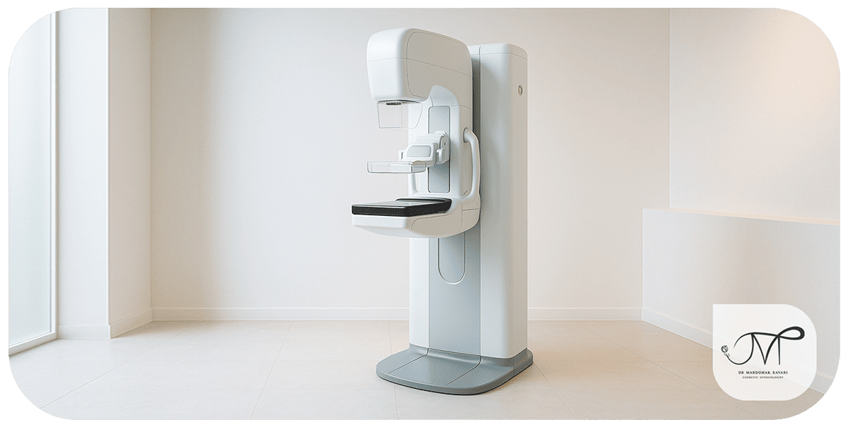
- Mammography: from age 40 (annually or every two years, based on doctor’s advice). High-risk women may start earlier and may need breast MRI.
- Breast awareness: self-checks aren’t perfect but help you notice changes earlier.
2) Clinical evaluation
Medical history and physical exam: checking both breasts, underarms, skin, and nipples.
3) Diagnostic imaging
- Diagnostic mammography and ultrasound to see if a lump is solid or fluid-filled.
- MRI in special cases (dense breasts, surgical planning).
4) Biopsy (the key to diagnosis)
Usually done with a core needle biopsy. The report shows the type of cells and receptor status (ER/PR/HER2).
5) Staging the cancer
Based on tumor size and spread to lymph nodes or other organs. In simpler terms: early, locally advanced, or metastatic. Staging determines the treatment plan.
Treatment options (benefits and notes)
Treatment combines different methods depending on stage and tumor biology. The goal is cure or long-term control while maintaining quality of life.
1) Surgery
Lumpectomy: removes only the lump and surrounding tissue; usually followed by radiation.
Advantage: preserves breast shape. Best for early stages.
- Mastectomy: removes the entire breast. In some cases reconstruction is possible immediately or later.
- Sentinel lymph node biopsy: checks for spread to nodes with minimal risk of lymphedema.
2) Radiation therapy
- Lowers risk of local recurrence (commonly after lumpectomy).
- Modern techniques (3D/IMRT) protect healthy tissue.
3) Chemotherapy
- Before surgery (neoadjuvant) to shrink the tumor, or after surgery (adjuvant) to reduce recurrence.
- Common side effects: fatigue, hair loss, nausea—but these are now more manageable.
4) Hormone therapy (for ER/PR+ tumors)
- Tamoxifen (before menopause) or aromatase inhibitors (after menopause).
- Oral, long-term treatment; requires monitoring for side effects.
5) Targeted therapies
- HER2 positive: trastuzumab, pertuzumab, and newer drugs have revolutionized outcomes.
- Advanced HR+: CDK4/6 inhibitors with hormone therapy provide longer control.
- BRCA mutations: PARP inhibitors may help.
6) Immunotherapy
For selected triple-negative cases (e.g., PD-L1 positive), combined with chemotherapy.
7) Supportive care and rehabilitation
Managing fatigue, nausea, sexual health, fertility planning, bone health, lymphedema prevention, and psychological support are essential parts of treatment.
Life after treatment: follow-up and lifestyle
- Regular check-ups (exams and imaging as advised).
- Physical activity, healthy weight, limit alcohol, avoid smoking.
- Mental health care; family and support groups are very helpful.
- Fertility and pregnancy planning should be discussed with the medical team.
Prevention and self-care

There’s no guaranteed prevention, but risk can be lowered by:
- Regular screening with mammography based on age and risk.
- Weekly exercise (at least 150 minutes).
- Maintaining a healthy weight.
- Limiting alcohol, avoiding smoking.
- Mediterranean-style diet: fruits, vegetables, whole grains, legumes, nuts, fish; less saturated fat and processed meat.
- Breastfeeding, if possible, slightly reduces risk.
- For high-risk women: genetic counseling, yearly MRI, preventive medication (e.g., tamoxifen), or in rare cases preventive surgery—always guided by specialists.
Clinical examples
Case 1 – Early detection with mammography:
A 46-year-old woman with no symptoms had a suspicious area on her routine mammogram. Biopsy showed early ductal carcinoma in situ. She underwent lumpectomy and radiation and returned to daily life. Screening made the difference.
Case 2 – HER2 positive with targeted therapy:
A 52-year-old woman had a 2 cm lump and one affected lymph node. Biopsy showed HER2 positivity. She received neoadjuvant chemotherapy with trastuzumab and pertuzumab, followed by lumpectomy and continued targeted therapy. She is now disease-free and active.
Case 3 – Triple negative with immunotherapy:
A 41-year-old woman presented with a rapidly growing lump. Diagnosed with TNBC, she received combined chemotherapy and immunotherapy before surgery, then surgery and radiation. She achieved a complete pathological response and returned to her normal lifestyle with regular exercise.
Conclusion
Breast cancer is common but manageable. The key lies in early detection and treatment tailored to the tumor’s biological type. If you notice any change, don’t delay.
Regular screening, a healthy lifestyle, and professional follow-up form the three pillars of breast health. In many cases, women visit their doctor due to a lump or change in the breast and worry about cancer, while the cause may actually be benign conditions such as breast cysts or fibroadenomas.
Both are typically noncancerous and cause noticeable changes in breast texture, but unlike cancer, they do not invade surrounding tissues. However, the distinction can only be made through clinical examination, imaging, and, if needed, biopsy.
Understanding these differences helps reduce patient anxiety and prevents unnecessary treatments. In some cases, mastitis (breast inflammation) can also mimic cancer with symptoms like redness, pain, or swelling.
This similarity highlights the importance of accurate evaluation — because a misdiagnosis can delay essential care. Therefore, any change in the breast — whether a lump, pain, or skin alteration — should be carefully assessed through specialist consultation and diagnostic imaging.
Proper differentiation between benign and malignant lesions ensures the right treatment at the right time, protecting both health and peace of mind.
Similar Articles

Don’t delay a proper breast lump evaluation!
Fill out the consultation form now and our assistant will contact you shortly.
 FAQ
FAQ
No comments have been posted.
Your email address will not be published. Required fields are marked *
Share:


